Green Body Analysis

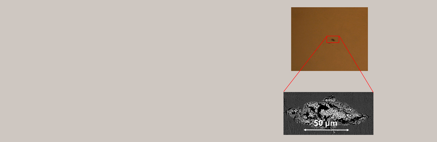
In order to achieve the desired product quality during thermal treatment, the green bodies must already possess suitable properties. In general, it is necessary for the green parts to be as homogeneous as possible on the micro, meso, and macro scales. The reasons for this are as follows:

  • Microscale: The individual particles must be arranged as uniformly as possible. Otherwise, there is a risk of preferential sintering of neighboring particles and premature grain growth.
  • Mesoscale: In sintered parts of this size, structural defects such as inclusions, large pores, or cracks are often present, which control the fracture behavior. Even a few structural defects per cubic centimeter lead to a deterioration in strength and reliability.
  • Macroscale: The density must be as constant as possible throughout the component, otherwise there will be uneven shrinkage and distortion during sintering. Distortion is associated with a loss of quality or an increased need for post-processing.

A careful assessment of the green body quality significantly reduces the complexity in the development of powder metallurgical manufacturing processes. Optimization steps in the areas of raw material selection, mass preparation, and shaping can then be largely separated from the optimization of thermal treatment and final processing.

Microscale
Inclosure in an oxide ceramic
© Fraunhofer Center HTL
Inclosure in an oxide ceramic, identified with immersion method and analysed with a SEM-micrograph (Scanning Electron Microscope)

The particle sizes of most green bodies are in the range of 0.1-100 µm. In this size range, scanning electron microscopy (SEM) is the most suitable method for imaging the structures. Flat sections through the microstructure are required for the quantitative assessment of homogeneity. These are produced in green bodies using a special almost artifact-free ion beam process called Cross Section Polishing (CSP). Subsequently, the green samples are imaged in the SEM with high contrast. An automated assessment of the homogeneity of the microstructure is performed using variance analysis with software developed specifically at Fraunhofer Center HTL.

Mesoscale
Computed tomography
© Fraunhofer Center HTL
Computed tomography at the HTL

Computed tomography (CT) is well suited for the examination of structural defects in green samples on a size scale above about 20 µm. This provides a 3D image of the green body with a resolution of up to a few micrometers. Software developed specifically at the HTL can also perform variance analysis to assess structural homogeneity using CT. Alternatively, a method has been developed at the HTL for ceramic green bodies, which can be used in production, to detect structural defects. In this method, the green parts are debinded and then soaked with an immersion solution that has the same refractive index as the ceramic. The samples become transparent and defects can be detected in the light microscope. The immersion method can be used with little effort to check the quality of green bodies once a suitable immersion solution has been found. However, it requires the use of exhaust systems, as the organic solvents used are harmful to health.

The defects found in the green bodies can be exposed using special target preparation methods and then analyzed in the SEM using energy-dispersive X-ray analysis (EDX). The origin of impurities can be narrowed down from the composition and/or shape of the defects.

Macroscale
Measurement of drying shrinkage
© Fraunhofer Center HTL
TOM_air: Measurement of drying shrinkage

In principle, there are many measurement methods available for analyzing the porosity or density distribution in green bodies:

  • The green bodies can be divided into small samples and their density can be determined geometrically or by Archimedes method.
  • Using X-ray techniques (transmission or computed tomography), the density of green bodies can be determined spatially resolved.
  • The sintering distortion can be measured and indirectly used to determine the density distribution in the green body.

The Archimedean or geometric measurement of the density of small samples taken from the green body requires only a small investment, but a high amount of effort. The accuracy of the density measurement is limited to approximately ±0.5% pore content, which is often insufficient for near-net-shape sintering. X-ray methods are much more elaborate in terms of equipment. They can provide important information on the binder and porosity distribution within the component. However, X-ray methods have limited significance in the edge region of the green parts, where particularly high density gradients are expected, because scattering effects occur. The most accurate method for detecting porosity gradients on a macroscopic scale is in-situ measurement of the sintering distortion. Samples must be taken from suitable locations in the green body. Small components can also be examined completely. The Thermo-Optical Measurement (TOM) method of the HTL then measures the shrinkage of the samples during sintering with high spatial resolution. The porosity in the green state is calculated from the shrinkage with an accuracy better than ±0.2%.

Service Offering:

  • Holistic evaluation of green body quality on micro, meso, and macro scales
  • Improvement of green body homogeneity through support in raw material selection, mass preparation, and shaping
  • Investigations into the influence of inhomogeneities on debinding and sintering behavior as well as component properties

You might also be interested in:

ICME

At Fraunhofer Center HTL, essential building blocks for integrated, computer-aided material development are available.

Effects of Defects

At Fraunhofer Center HTL, methods are being developed to quantitatively assess the effects of material defects on the reliability of components.

Product Control

Here you can find our flyer: